Amelanotic Melanoma
What is a Amelanotic Melanoma?
Amelanotic melanoma is a type of skin cancer in which the cells do not make melanin. They can be pink, red, purple or of normal skin color, hence difficult to recognise. It has an asymmetrical shape, and an irregular faintly pigmented border. Their atypical appearance leads to delay in diagnosis, the prognosis is poor and the rate of recurrence is high.
‘Amelanotic’ means without the usual brownish pigment produced by most pigment cells. Amelanosis is often a sign of tumour aggression and genetic diversity within the melanoma - in that the cells are generally so abnormal they no longer perform even the basic function pigment cells perform - make pigment. Such lesions often have a worse outcome (prognosis) than most other forms of melanoma.
Breslow Depth
Breslow refers to the skin depth of the melanoma. The Breslow Stage is used as a prognosis factor as it ranks how deeply the tumor cells have invaded. Breslow's depth was divided into 5 stages.
Early Breslow Amelanotic Melanoma Case Study
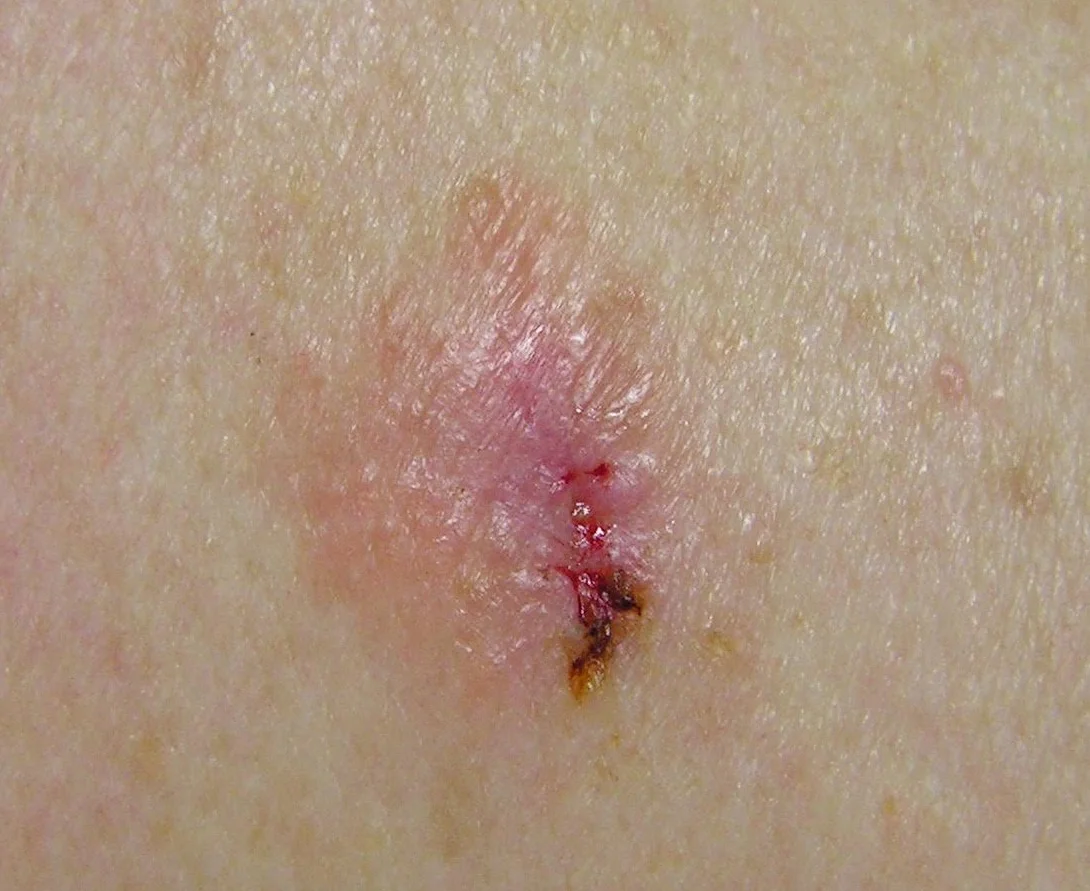
Amelanotic Skin Cancer Picture
Presentation
This is an early (Breslow 0.2mm) Amelanotic Melanoma appearing on the chest of a 47 year old female. It displays mixed colours of white, light and dark pink. The yellow-brown sections at the lower part of the lesion represent visible ulceration and associated scabbing.
Treatment
The lesion was widely resected and Sentinel Nodes were not assessed.
Prognosis
The patient is alive and well.
Small Amelanotic Melanoma Case Study
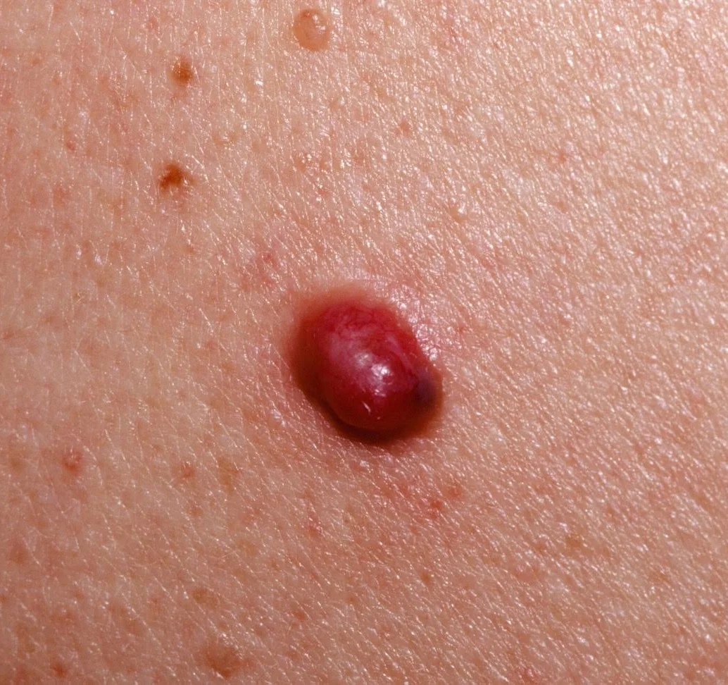
Small Amelanotic Melanoma Skin Cancer Picture
Presentation
A small (Beslow 0.8mm) Amelanotic Melanoma on the back of a 31 year old male. While the melanoma displays fronding of brown pigment at its lower pole, it is predominantly lacking pigment and so is referred to as Amelanotic.
A very small pearlescent (pearly coloured) Nodular Basal Cell Carcinoma can be seen at the top of the image.
Treatment
This lesion was widely resected and a Sentinel Lymph Node Biopsy was not performed.
Prognosis
The patient is alive and well but has gone on to develop several other skin cancers and precancerous lesions that have required further treatment.
Small Amelanotic Melanoma Case Study
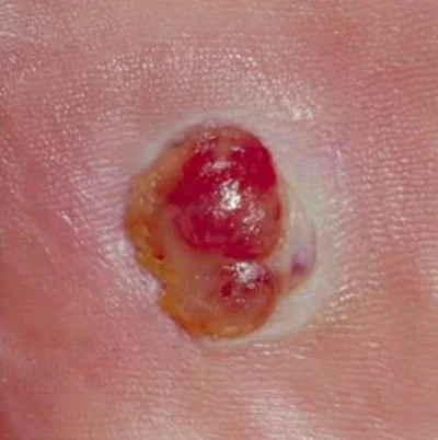
Medium Amelanotic Melanoma Skin Cancer Picture
Presentation
A (Breslow 1.2mm) Amelanotic Melanoma on the sole of the foot of a 24 year old female.
Treatment
This lesion was widely resected but Sentinel Node Lymph Biopsy proved positive.
An inguinal nodal clearance was performed. Tumours were found in 3 out of the 24 nodes and were removed.
Prognosis
The patient was disease free for another 2 years at which point brain metastases were detected on CT scanning performed to investigate complaints of headaches. The patient succumbed to the effects of widespread metastatic melanoma 3 months later.